Measuring mitochondrial mass as an indicator of cellular health and functional autophagy |  |
The phosphatidylinositol 3-kinase (PI3K) signaling pathway governs cellular functions, including cell cycle progression, metabolism, growth, and plays a role in autophagy. Two recent papers studying the role of PI3K in autophagy and amyloidogenesis examine mitochondrial mass as an indicator of cellular health.
In a publication in Science Advances, Tang et al. studied the effect of heat shock factor 1 (HSF1) activation in the PI3K/AKT signaling pathway through Ser230 phosphorylation. PI3K/AKT hyperactivation drives excessive protein translation and evokes amyloidogenesis. HSF1 neutralizes amyloid oligomers and thus protects the mitochondrial proteome’s stability to prevent apoptosis by shielding heat shock protein 60 (HSP60) from direct assault by amyloid oligomers. HSP60 is an essential mitochondrial chaperone that is required for proper folding and import of proteins into the mitochondria. Unregulated amyloidogenesis targets HSP60 and results in mitochondrial damage, apoptosis, and mitophagy. The decrease in mitochondrial mass, measured by flow cytometry of MitoView™ Green-stained astrocytes, confirms that the absence of HSF1 results in destabilization of HSP60. The increased apoptosis, measured by TUNEL and caspase 3 assay, further corroborates the protective effect of HSF1 on HSP60 in astrocytes from Hsf1 knockout mice.
In another article published in Cellular Molecular Immunology, Yang et al. studied the role of the PIK3C3 subunit of PI3K in the inflammatory functions of myeloid cells in the context of autoimmune encephalomyelitis. The PIK3C3 subunit of PI3K is a key early player in autophagy, promoting the degradation of cytoplasmic proteins and damaged organelles during immune cell response to various stimuli. The authors measured autophagy-related functions such as mitochondrial mass in myeloid cell Pik3c3-deficient mice to ascertain the role of PIK3C3 in inflammatory processes of myeloid cells. An increase in mitochondrial mass measured by cytometric bead array of MitoView™ Green-stained cells pointed to impaired autophagy. Annexin V staining of macrophages from the peritoneal cavity of Pik3c3 deficient mice confirmed defective uptake of apoptotic cells.
MitoView™ & Other Mitochondrial Dyes | ||
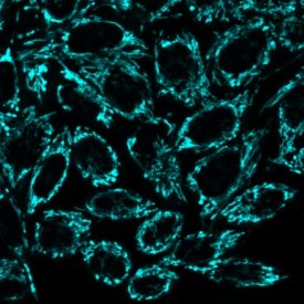
MitoView™ Blue |
Potential-independent MitoView™ Green dye |
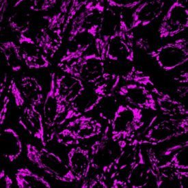
MitoView™ 633 |
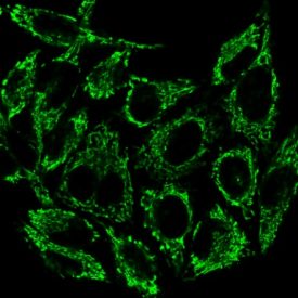
MitoView™ Dyes In metabolically active cells, mitochondria produce a membrane potential by maintaining a proton gradient across the inner and outer membranes. Loss of mitochondrial membrane potential is a hallmark for apoptosis. Biotium’s MitoView™ mitochondrial dyes are available in a variety of colors. MitoView™ Blue and MitoView™ 633 dyes are membrane potential-sensitive stains for mitochondria in mammalian cells and yeast. These dyes stain mitochondria with intact membrane potential in live cells. Staining is lost when mitochondria become depolarized during cell death, allowing monitoring of cell viability. We also offer MitoView™ Green, a membrane-potential independent mitochondrial dye that can be used to image mitochondria following mitochondrial depolarization, or after fixation. | JC-1 and Other Classic Mitochondrial Membrane Potential Dyes In healthy cells, JC-1 dye aggregates in mitochondria as a function of membrane potential, resulting in red fluorescence with brightness proportional to the membrane potential. Conversely, in apoptotic and necrotic cells with diminished mitochondrial membrane potential, JC-1 exists in a green fluorescent monomeric form in the cytosol, allowing of cell viability to be assessed by measuring the ratio of red to green fluorescence by flow cytometry or fluorescence microplate reader. |